Fig. 1
Chapter 1
WHAT IS MYOPIA?
The word myopia comes from a Greek word meaning closed eyes, and apparently referred to the squinting that nearsighted people resort to in order to see more clearly. Simply stated, myopia is a refractive error in which images come to a focus in front of the retina rather than on the retina, resulting in the inability to see distant objects clearly. To have a full understanding of what this means, one must first learn about the various parts of the normal eye and how they function. Figure 1 shows the principal parts of the eye.
Fig. 1
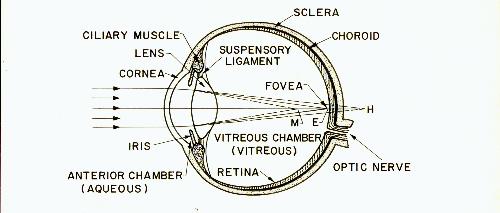
Parts of the eye. The light rays entering this eye are shown parallel to each other, as they would be if coming from a distant object. As the rays of light pass through the cornea or front surface of the eye, they are refracted or bent so that they begin to converge or approach each other.
The iris is the part of the eye that gives it its color, and the opening in the iris through which the light rays pass is called the pupil. If the iris contracts (as in bright light), the pupil gets smaller and reduces the amount of light that can continue further into the eye.
The light rays next pass through the lens where they are again bent and made to converge even more. If the rays have been bent just right, they will all come together (or focus) on the retina. The small central spot on the retina where we get the sharpest vision is called the fovea. Only in the fovea are the receptor cells packed close enough and appropriately organized to make a high degree of visual acuity possible.
This diagram shows what happens in the normal eye when it is in its relaxed condition, looking into the distance. This normal eye, in which distant rays come to a focus on the retina (at E), is said to be EMMETROPIC (pronounced em-e-trah'-pic). If, under the same set of conditions, the parallel rays would not come to a focus unless they were projected behind the retina (at H), the eye is said to be farsighted or HYPEROPIC (pronounced hy-per-ah'-pic). The words HYPEROPIC and HYPERMETROPIC are both in use but mean the same thing. If, under the same set of conditions, the parallel rays come to a focus in front of the retina (at M), the eye is said to be nearsighted or MYOPIC (pronounced my-ah'-pic). It is easy to see from this why the myopic eye is considered to be too long in relation to the parts of the eye that bend the light - the cornea and the lens. The words "myopia", "nearsightedness" and "shortsightedness" mean the same thing.
The anterior chamber contains a clear liquid called aqueous fluid or merely aqueous. The vitreous chamber contains a clear jelly-like substance called the vitreous or vitreous body.
The retina is only the innermost layer at the rear of the eye. The middle layer is called the choroid and the outermost layer is called the sclera. The choroid supplies the retina with blood and forms a dark lining inside the eye. These three layers are called the coats or tunics of the eye. The sclera becomes the cornea where it extends around the front of the eye.
The light that reaches the retina causes nerve impulses to be transmitted to the brain via the optic nerve.
The lens is made of flexible material and its shape can be changed by the ciliary muscle that surrounds it. This is what happens when the eye changes its focus. The ciliary muscle and lens are supported by the suspensory ligament.
Focusing. Figure 1 represents the normal eye in its relaxed condition looking at a distant object with the rays converging on the retina. We will next examine the changes which take place when that same eye looks at a near object.
Take a pencil in each hand and hold your arms straight out in front of you so that the pencils are the same distance from your eyes but are some distance apart. Now look at the top of the pencil in your right hand and focus on it so that it is seen clearly. The top of this pencil is now being projected onto the fovea of each eye. If you continue to focus your eyes on the right pencil, you will notice that the left pencil is not seen clearly. The reason for this is that the left-hand image is being projected onto a part of the retina that is much less sensitive than the fovea. It is not that the left pencil is out of focus. It is actually in focus, since it is the same distance from the eyes as the right pencil.
For the second part of this experiment, lay down the left pencil but continue to focus on the right pencil with only one eye. Close the other eye. You will notice that although the pencil is clear, objects closer or farther away than the pencil are not clear, even though they are directly in your line of sight and are consequently being projected toward the fovea of your eye. These objects are blurred because they are out of focus. Individual points on these objects are reaching the fovea not as individual points but as a whole circle of points, giving a blurred image.
If you open both eyes when doing this, you will notice that you can actually see two images of all objects closer or father away than the pencil. Since these objects are out of focus, they can easily be suppressed by the brain and the double images are not disturbing.
Now let us go on to another aspect of the focusing process. We can only see an object if it is giving off light. This can be light that is generated by the object itself, or it can be light that originates elsewhere but is reflected from the object. The sun is a good example of an object that can be seen by the light that it itself generates. The moon is a good example of an object that can be seen only by the light that it reflects (light which originally came from the sun).
Suppose we are looking at a small point on an object that is giving off light. As shown in figure 2, light rays are going off in all directions from this point source, but the only rays we see are the ones that pass through the lens.
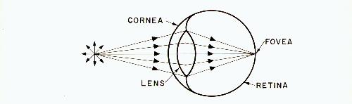
Fig. 2
Naturally, light does not really travel in individual rays like this, but it is more comprehensible to represent light in this manner.
In figure 2, the point source is shown quite close to the eye, and as the rays enter the eye they are bent properly so that they come to a point focus on the retina at the fovea. For simplicity, the bending of the light rays at the cornea is not shown in these diagrams. Such a bundle of light rays is sometimes called a pencil of light. All other points on the viewed object are giving off pencils of light that enter the pupil at different angles and come to a focus on different parts of the retina. In this manner, we see the entire object at one time. Since the eyes are capable of making very rapid movements, they can look at many different points on the object in rapid succession, giving us the impression that we see all points on the object with equal clarity.
Notice that the rays that enter the eye in figure 2 are getting farther away from each other as they approach the eye. Such rays are called diverging rays. In fact, all of the light given off by the point source consists of diverging rays. As the rays pass through the cornea and lens of the eye, they are bent so that they begin to come together again. In other words, they have become converging rays and will come together again at one point.
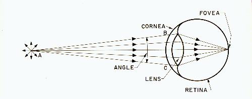
Fig. 3
In figure 3, the point source has been moved farther away from the eye. The rays that can enter the lens are less diverging than the corresponding rays in figure 2. In other words, the angle formed by the rays AB and AC has become smaller. As the point A is moved farther and farther away from the eye, the angle between these rays becomes smaller and smaller until, for all practical purposes, the rays entering the eye can be considered parallel. Therefore, figure 4 is a representation of the parallel light rays coming from a distant object.
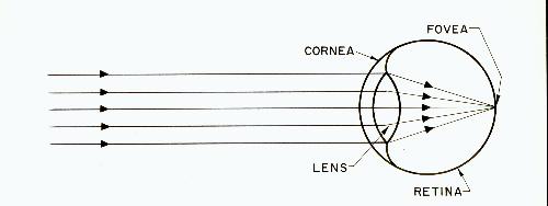
Fig. 4
Actually, since the pupil is so small, any object that is more than about two meters (six feet) from the eyes can be considered a distant object, at least from the standpoint of focusing the eyes. It is for this reason that distance vision can be tested by merely reading a chart on the opposite wall of the room. It is not necessary to look at something far in the distance.
The point to be remembered from the above is that the eye receives parallel rays from points on distant objects and diverging rays from points on near objects. This means that the eye has to adapt its focus to the distance of the viewed object.
Before explaining how the eye focuses, we can review something that is perhaps more familiar - the focusing of a camera.
If you have ever taken a picture, you know that the camera must be focused for whatever distance is being used. If the object is far away, the camera is focused for infinity; if the object is close, the camera must be focused for something less than infinity.
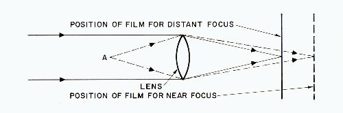
Fig. 5
Figure 5 shows a camera focused for infinity, with the solid lines indicating the parallel rays of light coming from a distant object and meeting on the film.
To take a picture of a near object at A, the distance between lens and film must be increased so that the rays of light can still meet on the film, as illustrated by the dotted lines.
Focusing of the human eye is done differently. The distance between the lens and retina is not changed. Instead, it is the lens itself that changes shape. The normal eye is at rest when looking into the distance and the light rays come to a focus as was shown in figure 4. Suppose that the eye now shifts its attention to a close object. If the eye were not able to change its focus, figure 6 shows what would occur. The cornea and lens of the eye do not bend the rays enough to focus them on the retina and the eye would see a blur. The eye solves this problem by a rapid focusing process that involves a change in the shape of the lens. Although the cornea bends the rays more than the lens, its shape remains fixed and it is not involved in this focusing process.
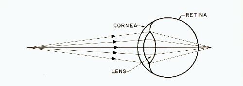
Fig. 6
The scientific term for this method of focusing on near objects is accommodation. By changing the shape of the lens an appropriate amount, the rays are made to bend more so that they again focus on the retina. Figure 7 shows the eye after it has accommodated for close vision.
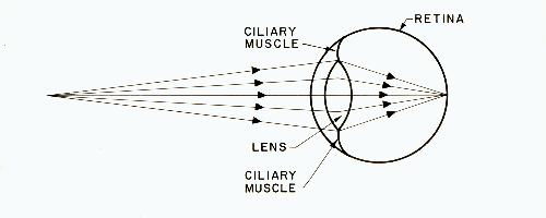
Fig. 7
Although accommodation, or focusing, usually takes place automatically without our being aware of it, it can also be accomplished by making a conscious effort to do so. In either case, it is the ciliary muscle, which surrounds the lens, that causes the lens to change shape.
Normally, the term "accommodation" is used to denote the active tightening of the ciliary muscle to focus for close work, and not to denote the relaxation of the ciliary muscle to focus for distance.
Referring back to figure 1, myopia was shown to be the condition where parallel rays from the distance always come to a focus in front of the retina. It is possible to state this problem in two ways:
Both these statements are really saying the same thing: There is an improper correlation between the length of the eye and the components that bend the light. The accepted myth is that this miscorrelation is inherited. The truth is that it is caused by the way we use our eyes.
The myopic eye can see clearly if the object is close enough because the diverging rays from such an object come to a focus further back in the eye.
The preceding diagrams dealt with how one point on a viewed object is focused to a corresponding point on the retina. Of course, in order to see the complete object, all points of the object must be projected onto the retina. Figure 8 shows how an arrow would be reproduced on the retina. Two points have been selected (the head and the tail of the arrow), and three lines from each point show the path of the light rays to the retina.
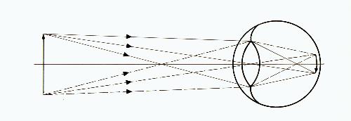
Fig. 8
The arrow is reproduced upside down on the retina but the brain interprets it as being upright. Remember that the entire arrow cannot be seen with equal clarity. If the axis of the eye is directed at the head of the arrow, then this point will fall on the fovea and be seen with maximum clarity. If the axis of the eye is directed at the tail of the arrow, then the tail will be seen with maximum clarity.
It should be obvious that myopia results merely from the improper optical properties of the eye and that it cannot be considered a disease. There are rare cases of congenital (present at birth) and of pathological (resulting from a diseased condition) myopia, but such cases are outside the scope of this book. Nearly all nearsighted people were born with normal vision and developed myopia because of the way they use their eyes. I prefer to call this type of myopia acquired myopia to indicate that it is something that develops in initially normal eyes. The expressions environmental myopia, simple myopia, school myopia and developmental myopia are also used to describe the same thing. Whatever it is called, it can be prevented.